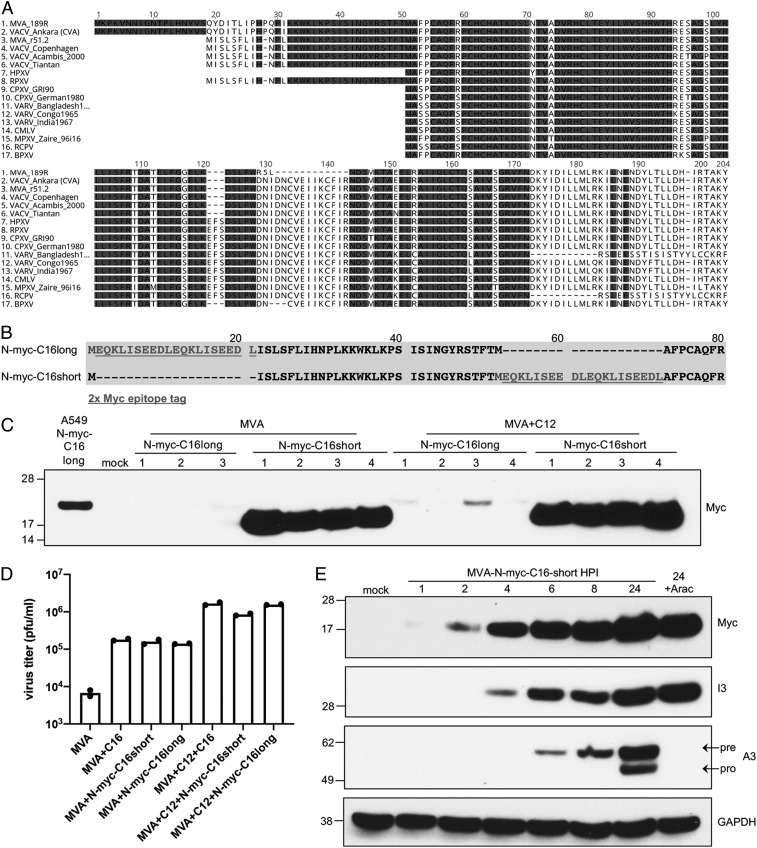
Fig. 2.
Sequence, expression, and activity of C16/B22 protein. (A) Multiple sequence alignment of C16L/B22R coding sequences from the indicated poxviruses. Only the B22R (189R) ORF of MVA is shown. For other orthopoxviruses, the two copies of the gene are identical, or only one copy is present. One hundred percent conserved residues are shaded. (B) Diagram showing placement of myc tag (underlined) before the first (N-myc-C16long) or second (N-myc-C16short) methionine. (C) A549 cells were mock-infected or infected with 5 pfu per cell of MVA+N-myc-C16long, MVA+N-myc-C16short, or the corresponding viruses that also express C12. C16long and C16short refer to placement of the Myc-tags after the Met at number 19 or 51 respectively, of the v51.2 C16 ORF in A. After 24 h, the cells were lysed and the proteins resolved by polyacrylamide gel electrophoresis. The C16 protein from A549 cells stably expressing codon-optimized C16 with a N-terminal Myc-tag after the first methionine regulated by the CMV promoter is shown in the first lane. Anti-myc-HRP was used for protein detection on Western blots. Numbers indicate individual clones of recombinant viruses. (D) A549 cells were infected with the indicated virus in duplicate at 0.01 pfu per cell for 48 h. Virus titers from each infection are shown as dots, and the bar represents the mean value. (E) A549 cells were mock-infected or infected with MVA-N-myc-C16short at 3 pfu per cell and after 1, 2, 4, 6, 8, and 24 h postinfection (hpi), analyzed by Western blotting. Numbers on the left are the masses in kDa and positions of marker proteins. The names on the right indicate the proteins recognized by antibodies. Pre and pro refer to precursor and processed proteins, respectively.

