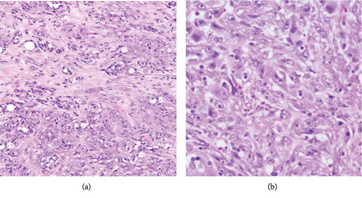
Figure 4.
Microscopic appearance of the tumor showing (a) complex, infiltrative, and poorly circumscribed cells with some cords and tubules (×10) and (b) carcinomatous proliferation formed by polygonal cells with eosinophilic and dense cytoplasm and an irregular hyperchromatic nucleus with multiple nucleoli and occasional presence of clear cells (×40).

