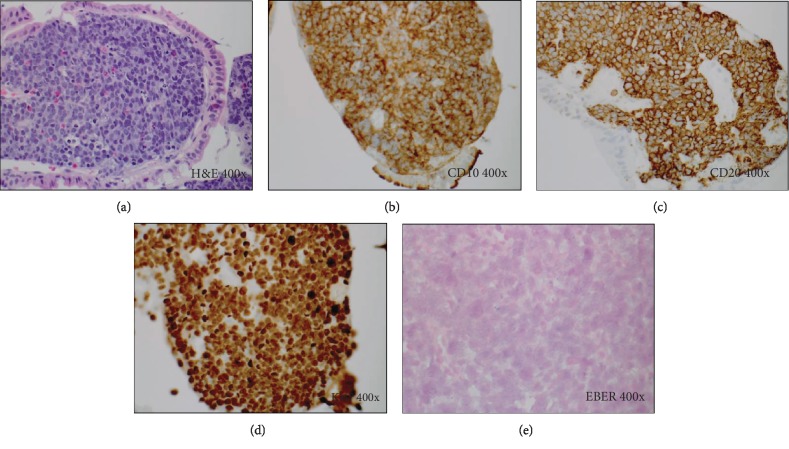
Figure 1.
Histology and special studies from the duodenal lesion in Case 1. (a) H&E stain (400x). Duodenal infiltrate composed of sheets of large monomorphic lymphoid cells exhibiting a classic “starry sky” appearance, characteristic of Burkitt lymphoma. (b, c) Immunohistochemical stains (400x) for CD10 (b) and CD20 (c) show strong diffuse positivity, while immunostains for BCL2 and TdT (not shown) are negative. (d) Ki67 immunostain (400x) demonstrates a very high proliferation rate of >90%. (e) In situ hybridization (400x) for Epstein-Barr encoded RNA (EBER) is negative.

