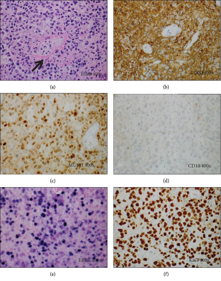
Figure 4.
Histology and special studies from the brain lesion in Case 5. (a) H&E stain (400x). Brain lesion showing an atypical lymphoid infiltrate composed of sheets of large lymphoid cells with abundant foci of single cell necrosis/apoptosis, often in a perivascular distribution (arrow). (b–d) Immunohistochemical stains (400x) demonstrate that the lymphoid infiltrate is composed of sheets of large B cells which are positive for CD20 (b), MUM1 (c), and BCL2 (not shown), but negative for CD10 (d). (e) The tumor cells are positive for EBER (400x) by in situ hybridization. (f) Ki67 immunostain shows a high proliferative rate of 90%.

