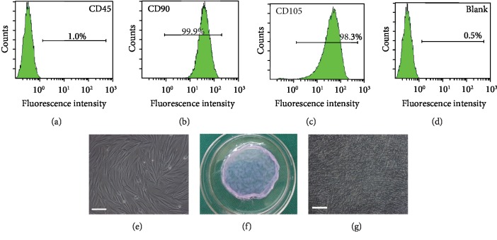
Figure 1.
Canine ADSC culture and ADSC sheet formation. (a–d) ADSCs were negative for hematopoietic marker CD45 and were strongly positive for MSC-related markers CD90 and CD105. The unlabeled cells were used as the blank control. (e) The primary cultured ADSCs were isolated from fat tissue of beagle dogs (scale bars: 100 μm). (f) ADSC sheets were obtained after 21 days of continuous cell culture. (g) ADSC sheets observed by inverted phase contrast microscopy (×100) and cell tight junction and surrounded by abundant ECM (scale bars: 100 μm).

