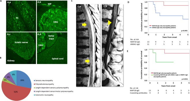
Figure 1.
Tissue expression, clinical presentations and outcomes of MAP1B neuropathy. Tissue-based immunofluorescence of serum from MAP1B neuropathy patient on rat tissue (A). Indirect immunofluorescence using serum of MAP1B-IgG seropositive patients demonstrating expression of protein in the cerebellum (Purkinje cells and dendrites in molecular layer (a) dorsal root ganglia and myenteric plexus (b), sciatic nerve (c), sympathetic tract and spinal cord (d)). Lack of rat kidney staining supports neural-specific tissue staining (c). Neuropathy phenotypes associated with MAP1B-IgG neuropathy phenotype (n=25, (B)). MRI of lumbar spine (C), demonstrating multifocal T2 hyperintensity involving the spinal cord (a) and gadolinium enhancement of lumbosacral roots (head arrow) on post-gadolinium T1 scan (b). Kaplan-Meier estimates of time to death from neurological symptom onset of neuropathy cases with MAP1B-IgG alone compared with those with MAP1B-IgG coexisting with ANNA1-IgG or CRMP5-IgG (D). Kaplan-Meier estimates of time to death from neurological symptom onset of neuropathy cases with MAP1B-IgG alone compared with those with ANNA1-IgG (E). ANNA1, anti-nuclear neuronal antibody type-1; CRMP5, collapsin response-mediator protein-5; DRG, dorsal root ganglia; MAP1B, microtubule-associated protein 1B; ML, molecular layer; MP, myenteric plexus; PC, Purkinje cell; Symp, sympathetic.

