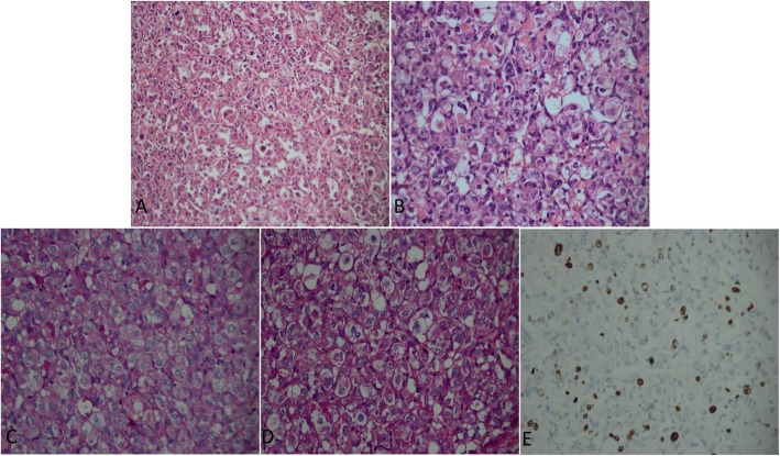
Fig. 3.
a Photomicrograph of the tumour section showing nests of large polygonal cells separated by thin fibrovascular septae (haematoxylin-eosin, original magnification × 200). b The cells are large, uniform, and epithelioid with fine, eosinophilic, granular cytoplasm and eccentrically located nuclei containing prominent nucleoli (haematoxylin-eosin, original magnification × 400). c PAS revealing characteristic diastase-resistant crystals that vary in size and shape within the cytoplasm of the tumour cells (PAS, original magnification × 400). d D-PAS-positive crystalline structures in tumour cells showing non-glycogen materials (D-PAS, original magnification × 400). e Ki67 immunostaining of tissue demonstrating strong nuclear immunoreactivity in some tumour cells (Ki67, original magnification × 400)

