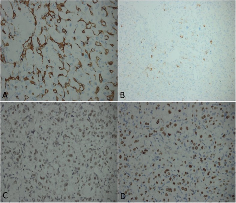
Fig. 4.

a CD34 immunostaining of specimens showing diffuse cytoplasmic immunoreactivity in tumour cells (CD34, original magnification × 400). b Desmin immunostaining of tumour cells revealing cytoplasmic positivity (Desmin, original magnification × 400). c INL-1 staining of tumour cells showing nuclear immunoreactivity (INL-1, original magnification × 400). d Some tumour cells of the specimen show nuclear immunoreactivity after TFE-3 staining (TFE-3, original magnification × 400)
