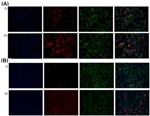
Figure 10.
Representative fluorescence microscopy images showing the uptake of liposomes by PAH-SMCs after (A) immediate and (B) 24 h of incubation of (i) fluorescent liposomes without CAR peptide and (II) fluorescent CAR-conjugated liposomes in cell culture media. Green, beta actin; blue, DAPI; red, rhodamine B-labeled liposomes. The rightmost panel shows the overlay.

