Figure 3.

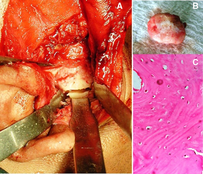
Intraoperative image showing exposure of lesion with preauricular incision and osteotomy cut placed for condylectomy (A), excised mass (B). Photomicrograph showing dense compact bone with osteocytes (×40 magnification) (C).

Intraoperative image showing exposure of lesion with preauricular incision and osteotomy cut placed for condylectomy (A), excised mass (B). Photomicrograph showing dense compact bone with osteocytes (×40 magnification) (C).