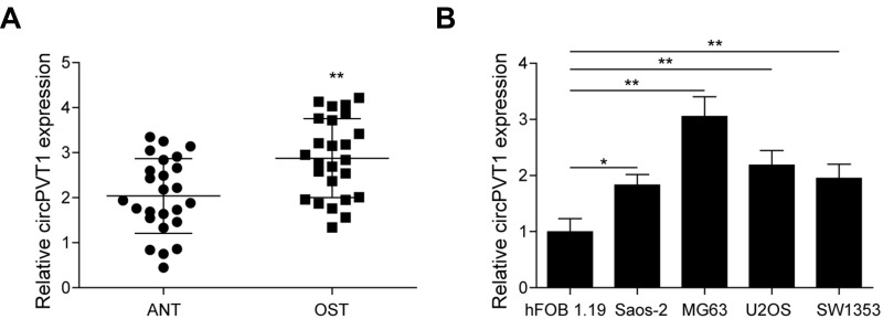
Figure 1.
The expression of circPVT1 in OS tissues and cell lines. (A) qRT-PCR analysis of circPVT1 in clinical OS and adjacent normal tissue samples. (B) qRT-PCR analysis of circPVT1 in hFOB1.19 cell line and four OS cell lines (Saos-2, MG63, U2OS and SW1353). The data were means ± SD of three independent assays, *P < 0.05; **P < 0.01.

