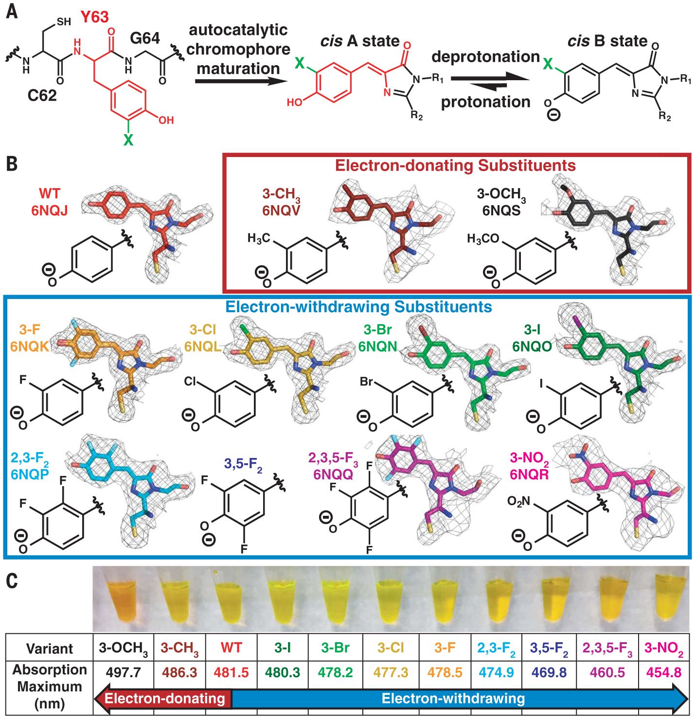
Fig. 2. Incorporation of electron-donating and electron-withdrawing substituents into the Dronpa2 chromophore.

(A) Scheme depicting incorporation of substituents (represented by a green “X”) through amber suppression of Y63 and chromophore maturation in Dronpa2 variants (C, Cys; Y, Tyr; G, Gly). (B) Dronpa2 amber suppression variants grouped by electron-donating and electron-withdrawing properties. The electron density maps (2mFo – DFc, 1σ) from solved x-ray structures (except 3,5-F2, which could not be crystallized; see supplementary text S2) show substituent orientation(s) (see fig. S2 for omit maps). Two conformations were necessary for modeling the chromophore of the 3-F variant (fig. S2). The legend of fig. S2 includes the identity of the monomer displayed for each variant. WT, wild type. (C) Image of purified proteins and their corresponding 77 K absorption peak maxima.
