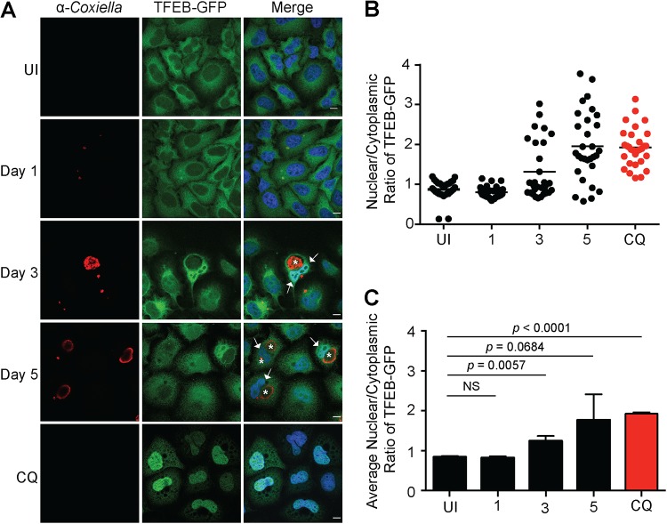
FIG 2.
Nuclear translocation of TFEB-GFP during C. burnetii infection of HeLa TFEB-GFP cells. (A) Representative confocal images of HeLa TFEB-GFP cells that were uninfected (UI); infected for 1, 3, or 5 days with C. burnetii NMII at an MOI of 100; or treated for 24 h with 50 μg/ml chloroquine (CQ). Following infection and/or treatment, cells were fixed and immunolabeled with anti-C. burnetii (red), and nuclei were stained with DAPI (blue). White arrows and asterisks denote TFEB nuclear translocation and CCVs, respectively. Bars, 10 μm. (B) Representative nuclear/cytoplasmic ratios of TFEB-GFP from 30 uninfected, infected, or treated cells at the time points indicated. (C) Average nuclear/cytoplasmic ratios of TFEB-GFP from three biological replicates. The means and SD for each time point are presented. An unpaired two-tailed t test was used to determine the statistical significance between samples. NS, not significant.

