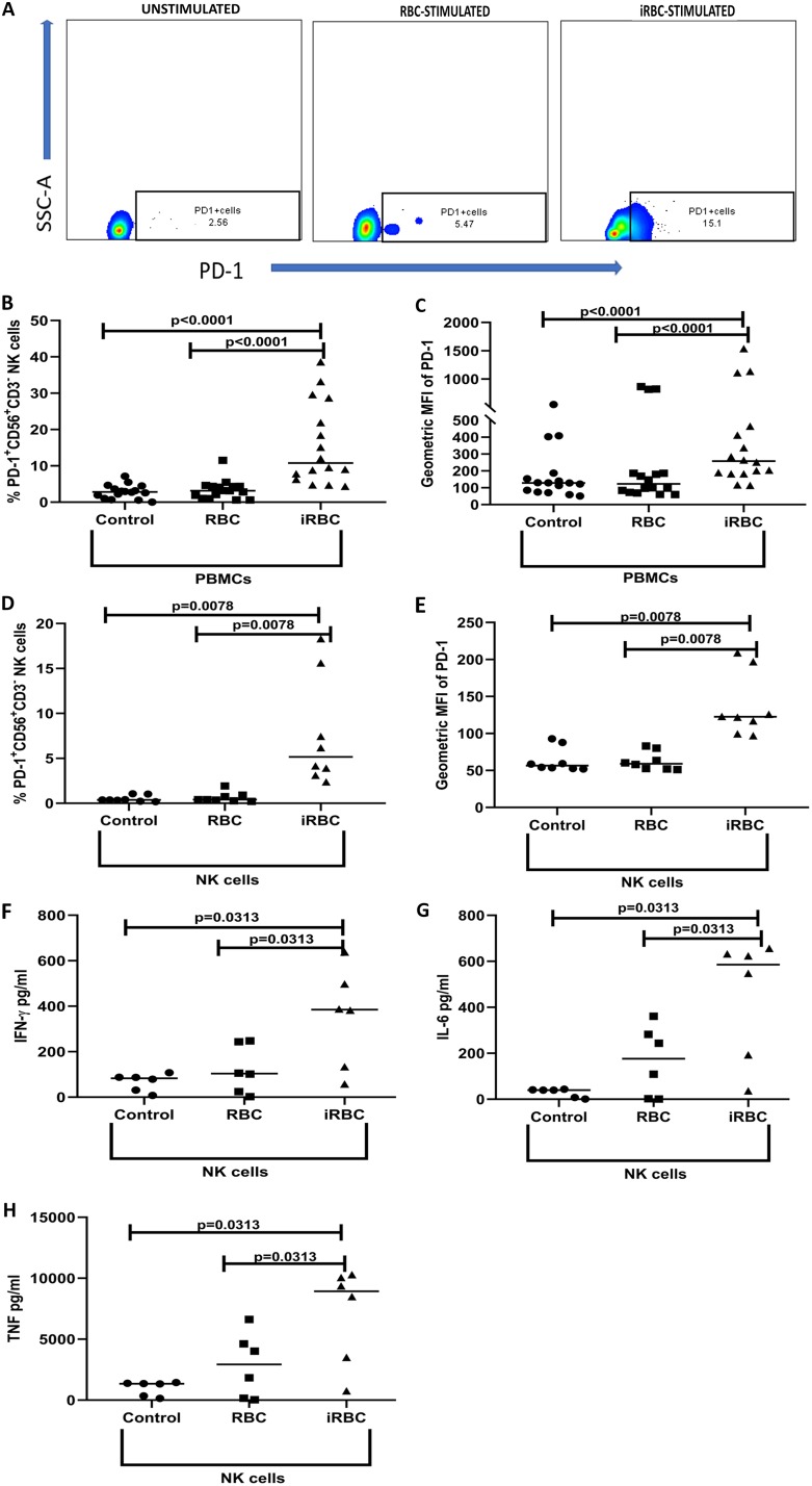
FIG 3.
NK cells of malaria-naive adults upregulate PD-1 following P. falciparum exposure in vitro. PBMCs (n = 16) or NK cells (n = 8) from malaria-naive U.S. adults were incubated at 37°C for 3 days with medium or with iRBCs or RBCs at a ratio of 1:30 (1 PBMC or NK cell to 30 iRBCs or RBCs) and analyzed by flow cytometry. Supernatants were removed at 24 h from the purified NK cell culture to quantify secreted cytokines (n = 6). (A) Representative flow cytometry plots show the gating strategy to identify PD-1+ CD3− CD56+ NK cells within FSC-SSC lymphocytes after gating on singlet live cells. (B) Percentage of total NK cells expressing PD-1. (C) MFI values of PD-1 on NK cells. (D) Percentage of purified NK cells expressing PD-1. (E) MFI values of PD-1 on purified NK cells. (F to H) Levels of IFN-γ (F), IL-6 (G), and TNF (H) in supernatants. P values were determined using the Wilcoxon signed rank test. Data are shown as medians (B to H).

