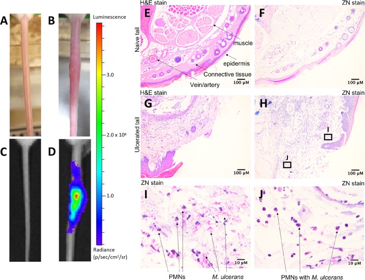
FIG 2.
Characterization of infection using a low-dose bioluminescent M. ulcerans strain. (A and B) Representative light camera images of tails from an uninfected BALB/c mouse (A) or at the point of ulceration (16 weeks) following intradermal inoculation with 20 CFU of bioluminescent M. ulcerans (B). (C and D) The same tails were visualized under an IVIS camera to detect and quantify bioluminescence intensity (as photons [p]/s). (E to H) Histological cross-section of an uninfected (E and F) or infected tail tissue (G and H) following hematoxylin & eosin (H&E) and Ziehl-Neelsen (ZN) staining. (I and J) Zoomed images of the regions indicated within the denoted boxes of panel H depict the presence of polymorphonuclear cells (PMNs) and acid-fast bacilli (ZN staining) within tissue.

