FIG 1.
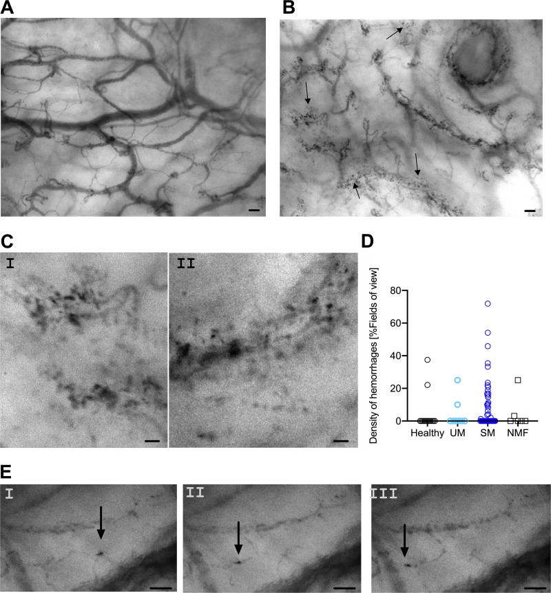
IDF imaging shows malaria-induced changes in the buccal microcirculation. (A) A still image of a healthy volunteer showing highly delineated blood vessels. (B) A still image from malaria-infected individuals with multiple hemorrhages. (A still image of a malaria-infected individual without hemorrhage is seen in Fig. S1 in the supplemental material.) Arrows in panel B denote perivascular hemorrhages. (C) Close-ups of representative hemorrhages; panel I is selected from the still shown in panel B, while panel II is a close-up from a different still image. (D) Density of perivascular hemorrhages as determined by IDF imaging. (E) Still images from a movie showing an infected erythrocyte sequestering in a capillary (Movie S1). Still images are temporally separated by 40 ms. Scale bars, 100 μm (A and B) and 50 μm (C and E).

