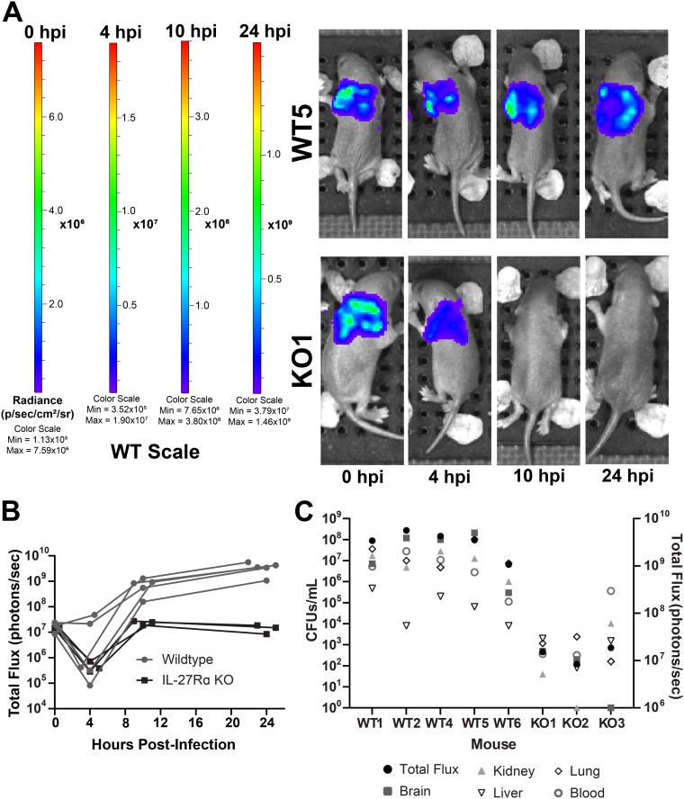
FIG 6.
Intravital longitudinal imaging of the influence of IL-27 during neonatal sepsis. Neonatal C57BL/6 (WT) and IL-27Rα−/− (KO) mice were subcutaneously inoculated with ∼2 × 10⁶ CFU/mouse of luciferase-expressing E. coli O1:K1:H7 or PBS as a control on day 4 of life in parallel. The neonatal pups were imaged longitudinally on an Ivis SpectrumCT at 0, 4, 10, and 24 h postinfection (hpi). Each mouse was tail tattooed for individual identification during imaging. Data are the result of an independent experiment (WT, n = 5; KO, n = 3) representative of two experiments with similar results. (A) Luminescent images of representative WT and KO mice at 0, 4, 10, and 24 hpi. The signal shown is on the WT scale. Colorimetric scale: low (minimum) signal is blue, intermediate signal is green, and high (maximum) signal is red. (B) Total luminescent flux in photons/second for individual mice at 0, 4, 10, and 24 hpi. (C) At 24 hpi, mice were sacrificed, and blood and peripheral tissues were collected for enumeration of bacteria by standard plate counts. Total luminescent flux (photons/second) and CFU/ml from each tissue for individually infected mice are shown.

