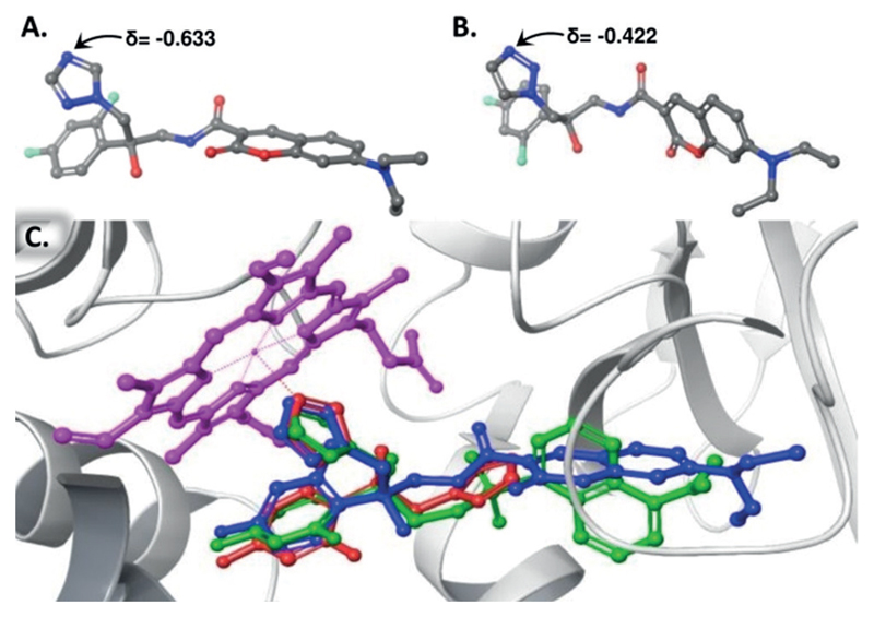
Figure 2.
A) Partial charge value of the N-4 of the 1,2,4-triazole-based 3. B) Partial charge value of the N-3 of the 1,2,3-triazole-based 5. The partial charges were assigned according to the OPLS3 force field calculations. C) Docked structure of the R-enantiomer of azole 2 (green), the R-enantiomer of azole 3 (blue), and FLC (red) on a crystal structure of S. cerevisae cytochrome P450DM (PDB ID: 4ZDY). Protein residues are shown as grey ribbons with the heme prosthetic group in magenta.

