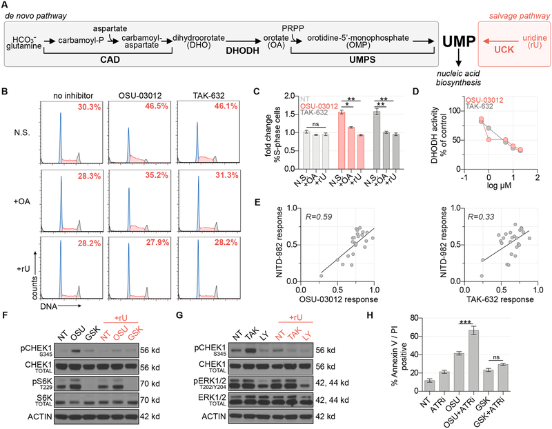
Figure 3 |. OSU-03012 and TAK-632 inhibit DHODH and activate the DNA replication stress response pathway.
(A) Schematic of UMP biosynthesis via the de novo and salvage pathways. (B) Propidium iodide cell cycle analysis of MIAPACA2 PDAC cells treated ±5 μM TAK-632 or ±5 μM OSU-03012 and supplemented with 50 μM orotate (OA) or 10 μM rU (N.S.: no supplement). Insert indicates % S-phase cells. (C) Summary of fold changes in S-phase cells from B (mean±SD; n=2; one-way ANOVA corrected for multiple comparisons by Bonferroni adjustment, ns: not significant; * P<0.05; ** P<0.01). (D) in vitro DHODH enzyme assay performed in the presence of OSU-03012 or TAK-632. (E) Correlation between DHODH inhibitor (1 μM NITD-982) and OSU-03012 (3.17 μM) or TAK-632 (3.17 μM) response across a panel of 25 PDAC cell lines determined using CTG following 72 h treatment. Response calculated as doubling time normalized proliferation inhibition. Pearson correlation coefficient is indicated. (F) Immunoblot analysis of MIAPACA2 cells treated ±1 μM PDK1 inhibitor GSK-2334470 (GSK) ±1 μM OSU-03012 (OSU) ±10 μM rU for 24 h. (G) Immunoblot analysis of MIAPACA2 cells treated ±10 μM RAF inhibitor LY3009120 (LY) ±10 μM TAK-632 (TAK) ±10 μM rU for 24 h. (H) Annexin V/PI flow cytometry analysis of MIAPACA2 PDAC cells treated ±1 μM OSU-03012 or 1 μM GSK-2334470 (GSK) ±500 nM VE-822 (ATRi) ±25 μM rU for 72 h (mean±SD; n=2; one-way ANOVA corrected for multiple comparisons by Bonferroni adjustment; ns: not significant; ** P<0.01; *** P<0.001).

