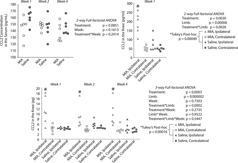
Figure 4. MIA-injected knees show elevated CCL-2 levels compared to controls and contralateral knees.
CCL2 levels in rat knee and in serum at 1, 2, and 4 weeks after MIA injection, as assessed by lavage, magnetic capture, or direct ELISA (serum). No statistically significant differences were identified between the CCL2 serum levels of MIA and saline injected animals (2-way ANOVA, left top panel). On the contrast, magnetic capture detected an increase of CCL2 in the MIA-injected knee relative to saline animals and relative to the contralateral knees within the same time point; time point was not a significant factor (3-way ANOVA, bottom panel). Lavage results confirm elevated CCL2 concentration in MIA-injected knee at the one-week time point (right top panel).

