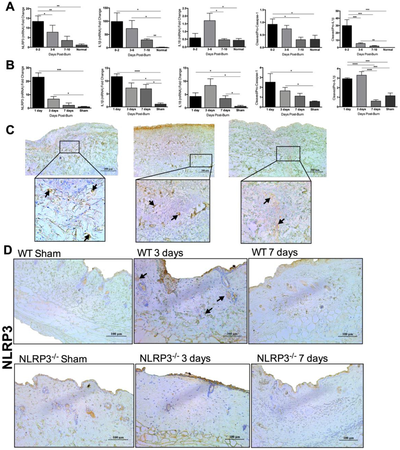
Figure 1.
NLRP3 is upregulated in human and murine skin after burn. (a) Time course of gene expression of NLRP3, IL1β and IL18 and protein expression of cleaved to pro-caspase-1 and cleaved to pro-IL1β at 0-10 days after burn compared to normal skin. (b) Gene expression of NLRP3, IL1β and IL18 and protein expression of cleaved to pro-caspase-1 and cleaved to pro-IL1β in murine skin after burn. (c) Immunohistochemical staining for NLRP3 at 0-2 days post-burn in human burn skin indicates more NLRP3 positive cells in the dermis. (d) Staining for NLRP3 at 3 and 7 days after burn in murine skin indicates more NLRP3 positive cells in the dermis of WT at 3 days. Areas of positive staining are marked with an arrow. Values are presented as mean ± standard error. Burn versus normal skin *p < 0.05; **p < 0.01; ***p<0.001.

