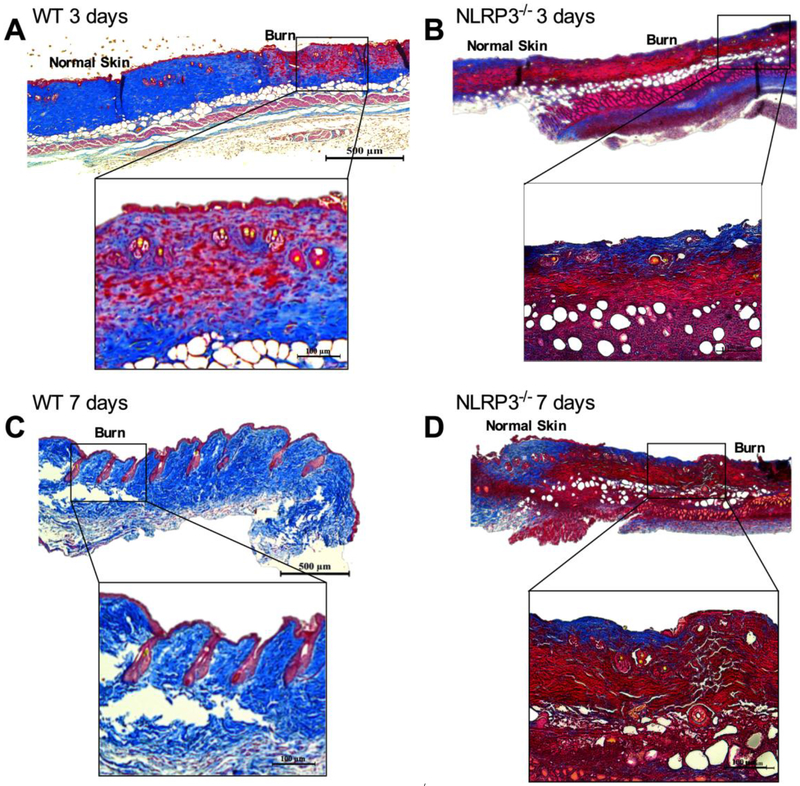
Figure 2.
Impaired wound healing in NLRP3−/−. Trichrome staining of excised burn wounds in WT at (a) 3 days and (c) 7 days exhibits greater dermal collagen deposition and keratinization compared to NLRP3−/− at (b) 3 days and (d) 7 days post-burn. “Normal skin” demarcates the edge of the wound, and “burn” denotes the wound.

