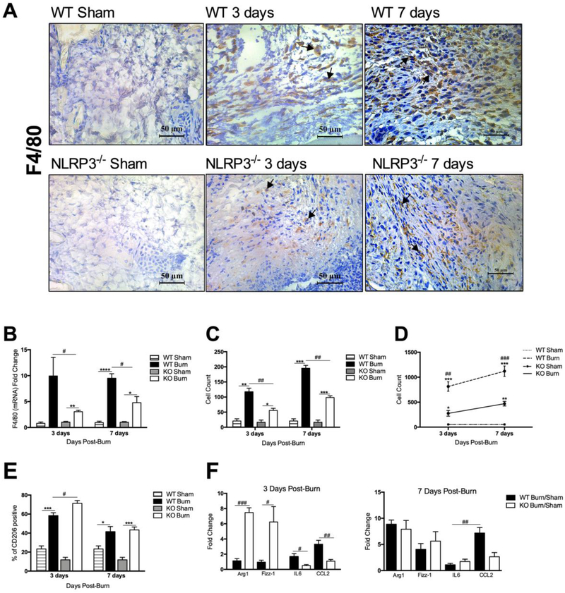
Figure 3.
Reduced macrophage proportion acutely after burn in NLRP3−/−. (a) F4/80 staining for macrophage infiltration in NLRP3−/− and WT mice shams, 3 days and 7 days after burn is corroborated by (b) PCR analysis, which indicates greater expression of F4/80 in WT at both time points. (c) F4/80-positive cell counts obtained at 40X magnification (n=3). (d) Flow cytometry for skin macrophage distribution shows greater macrophage infiltration in WT. (e) Flow cytometry analysis for CD206-positive anti-inflammatory macrophages indicates a greater percentage in NLRP3−/− at 3 days compared to WT. (f) Expression of pro- and anti-inflammatory markers at 3 days indicates a pro-inflammatory profile in WT and an anti-inflammatory profile in NLRP3−/−, with no significant differences in marker expression between WT and NLRP3−/− at 7 days. Values are presented as mean ± standard error. Burn versus sham *p < 0.05; **p < 0.01; ***p< 0.001, WT versus NLRP3−/− burn #p < 0.05; ##p < 0.01; ###p < 0.001.

