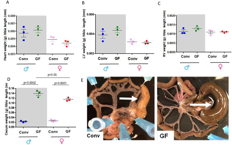
Figure 2:
Total heart (A), left (LV, B) and right (RV, C) ventricles and cecum (D) weights were measured and normalized by tibia length from 7–8 weeks old male and female Conv or GF mice. Number of animals used per group are = 3. Values are mean ± SEM. One-Way ANOVA and t-test. The images are representative pictures from the mesenteric bed of the male Conv and GF mice (E). Arrows show the cecum. Please note that the mesenteric bed from the GF mice presents an enlargement of the cecum. This is a typical characteristic for GF animals.

