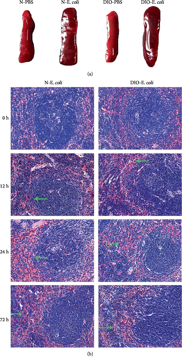
Figure 5.
E. coli-induced histological changes in normal and DIO mice. (a) Representative spleen anatomy changes following E. coli (12 h). (b) H&E staining of spleen sections was visualized via light microscopy to examine spleen architecture. Images were taken at 400x magnification. n = 8 mice per group. The arrow points to hyperemia.

