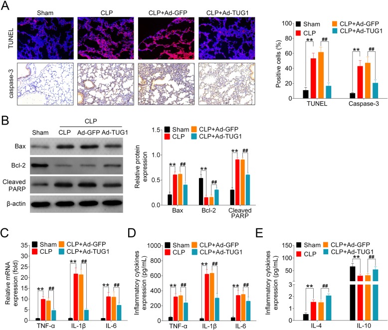
Fig. 2.
Impact of TUG1 upregulation on CLP-induced apoptosis and inflammation in mouse lung tissues. (a) Histological analysis of sectioned mouse lung tissue samples using TUNEL staining and immunohistochemistry for caspase-3 expression. Representative histological images (400× magnification) and the percentage of positively stained cells were shown. (b) The levels of Bax, Bcl-2, and cleaved PARP in the lung tissues of all mice were analyzed using Western blot. Representative plots (left) and bar charts (right) were shown. (c-d) The mRNA and proteins expressions of TNF-α, IL-1β and IL-6 in all animals were detected using qRT-PCR and ELISA, respectively. (e) The protein expressions of IL-4 and IL-10 in mouse lung tissues were measured using ELISA

