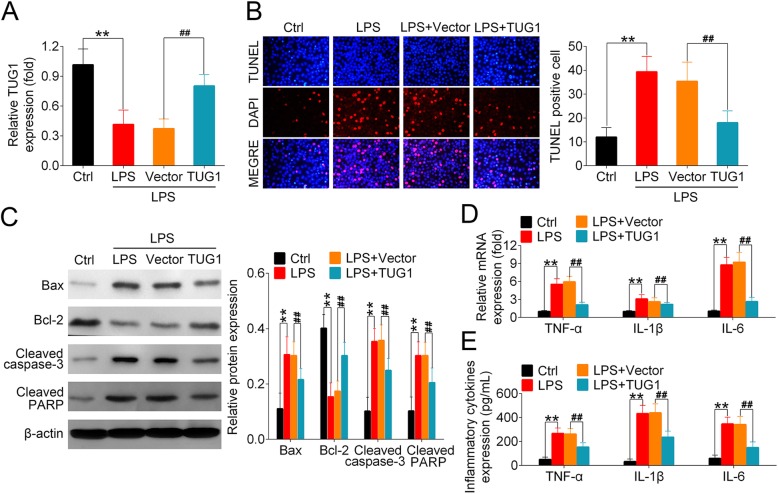
Fig. 3.
Overexpression of TUG1 mediated the levels of apoptotic markers and proinflammatory cytokines in LPS-treated PMVECs. PMVECs were divided into 4 groups: control (Ctrl), LPS, Vector, and TUG1. LPS, Vector, and TUG1 groups were cultured with serum-free medium containing nothing, control adenoviral vector, and adenoviral vector expressing TUG1, respectively, followed by the 6-h stimulation of 100 ng/mL LPS. Ctrl group remained untreated. (a) Relative expression of TUG1 were measured using qRT-PCR 6 h after LPS treatment. (b) TUNEL and DAPI staining were performed to detect apoptotic cell death in PMVECs. The number of TUNEL-positive cells were counted in six randomly selected fields per slide. (c) The expression of Bax, Bcl-2, cleaved caspase-3, and cleaved PARP in all groups of PMVECs were examined using Western blot. Representative plots (left) and bar charts (right) were shown. (d-e) The mRNA and proteins levels of TNF-α, IL-1β and IL-6 in PMVECs were assessed using qRT-PCR and ELISA, respectively

