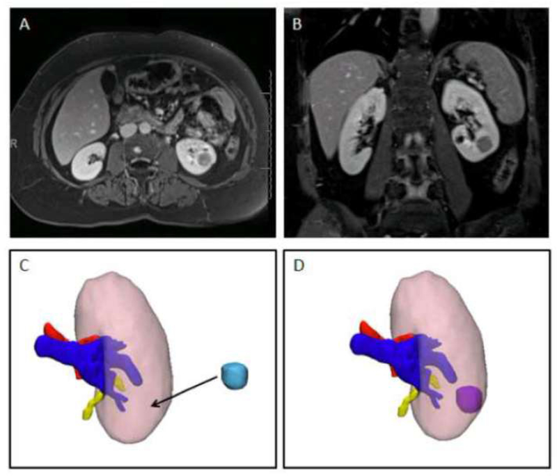
Figure 1.
Single case example showing A) axial and B) coronal images of a left endophytic renal mass. C) 3D kidney model with the kidney – pink, artery-red, vein- blue, and collecting system – yellow. The tumor (light blue) has been removed so the surgeon can translate the location where he believes it is after reviewing the images. D) 3D kidney tumor model with tumor in its correct location.

