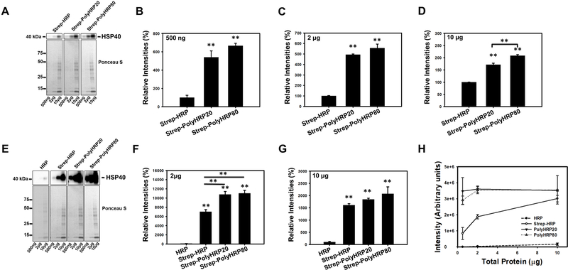
Figure 2.
Western Blots of rat heart lysates probed for heat shock protein 40 (HSP40) using Streptavidin-HRP (Strep-HRP) or Streptavidin-conjugated PolyHRPs (Strep-PolyHRP20, and Strep-PolyHRP80). (A) Figure shows representative unsaturated bands of HSP40 detected in 500ng, 2μg, and 10μg of heart lysates, and corresponding Ponceau S stained membranes. B-D shows the quantification of the HSP40 blots. Ponceau S was used for normalization of the Western blotting results. (B-D) Quantification of HSP40 using Strep-HRP, Strep-PolyHRP20, and Strep-PolyHRP80 on 500ng (B), 2μg (C), and 10μg (D) of heart lysate. (E) The upper figure shows representative blots of HSP40 detected in 500ng, 2μg, and 10μg of heart lysates. To detect the HSP40 using classical Western blotting methods together with the PolyHRP based methods the HSP40 detected using PolyHRPs had to be overexposed for the 2 and 10 μg of heart lysates. The lower figure shows Ponceau S staining which was used for normalization of the blots. Quantification of HSP40 using HRP, Strep-HRP, Strep-PolyHRP20, and Strep-PolyHRP80 on 2μg (F), and 10μg (G) of heart lysate with the bands using Strep-HRP, Strep-PolyHRP20, and Strep-PolyHRP80 all being saturated. (H) Graph showing the correlation between the amounts of heart lysate versus band intensity for HSP40. Values are means ± SE; (n = 3) per group. *P<0.05, **P<0.01.

