Fig. 1.
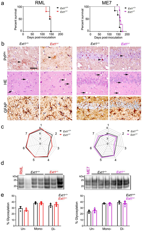
Ext1+/+ and Ext1+/− mice infected with subfibrillar prions show similar survival times, brain lesions, and biochemical properties. a A comparison of survival times revealed no differences in RML-infected Ext1+/− mice and a modest delay in ME7-infected Ext1+/− mice as compared to the Ext1+/+ mice. b Brain sections immunolabelled for PrP and GFAP, or stained with hematoxylin and eosin (HE), show indistinguishable prion aggregate distribution and morphology (arrowheads), spongiform degeneration (arrows), and astrogliosis in Ext1+/+ and Ext1+/− brains. c Lesion profiles of RML- and ME7-infected Ext1+/+ and Ext1+/− mice (1-dorsal medulla, 2-cerebellum, 3-hypothalamus, 4-medial thalamus, 5-hippocampus, 6-septum, and 7-cerebral cortex) are almost superimposable. d Electrophoretic mobility and e glycoprofiles of RML and ME7 strains in Ext1+/+ and Ext1+/− mice. The RML and ME7 inocula were loaded for comparison (first lane). *P< 0.05, Log-rank (Mantel-Cox) test (panel a). Cerebral cortex (RML) and hippocampus (ME7) shown in panel b. Scale bar = 50 μm (panel b). RML: n=6-7 mice/group; ME7: n=4-5 mice/group.

