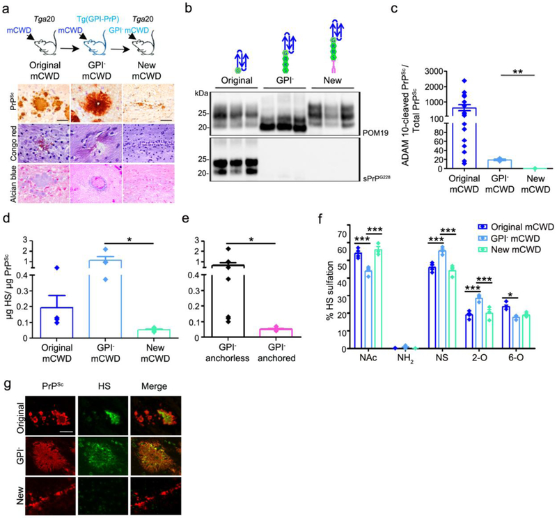
Fig. 5.
GPI-anchored mCWD prions do not form plaques and bind low levels of HS. a Schematic representation of mCWD inoculated into tga20 and GPI-anchorless PrPC expressing mice [tg(GPI-PrP)] with corresponding prion plaque morphology, Congo red, and Alcian blue staining of brain sections (corpus callosum). Note that the new mCWD prions do not bind Congo red or Alcian blue. b Western blots of PK-treated PrP labelled with anti-PrP POM19 (total PrP) or sPrPG228 (ADAM10-cleaved PrP) antibodies reveal ADAM10-cleaved PrP in the original mCWD but not the GPI-anchorless mCWD or the new mCWD–infected brain. c Ratios of ADAM10-cleaved : total PrPSc are higher in the original mCWD than in the GPI-anchorless mCWD or in the new mCWD-infected brains (original mCWD results are also shown in Fig. 4c). d The new mCWD binds less HS than the original mCWD or GPI-anchorless mCWD (original mCWD results are also shown in Fig. 4f). e A grouped comparison shows HS bound to GPI-anchorless prions (mCWD and GPI− mCWD, black bar) versus the new GPI-anchored mCWD (pink bar). f The HS bound to GPI-anchorless mCWD is more sulfated than the HS associated with ADAM10-cleaved mCWD and the new GPI-anchored mCWD, as it contains less unsulfated N-acetylated (NAc) HS and higher level of N-sulfated (NS) and 2-O sulfated (2-O) HS. g HS does not co-localize to the new mCWD aggregates (bottom panel). Scale bars = 100 μm for original and GPI− mCWD and 200 μm for new mCWD (panel a) and 25 μm (panel g). *P< 0.05, **P< 0.01 and ***P< 0.001, Wilcoxon rank sum test (panels c and e), one-way ANOVA with Tukey’s post test (panel d), and two-way ANOVA with Bonferroni’s post test (panel f).

