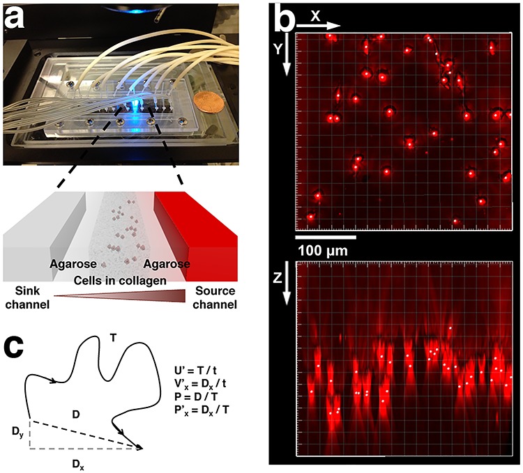
Figure 1.

A microfluidic setup for 3D tumor cell migration in CCL19 gradients. (a) Top view shows a photo of the microfluidic chip on a microscope stage. Each chip contains four identical three-channel devices. Zoom in view shows that each device contains three parallel channels with a width of 400 μm and a depth of 250 μm. The gaps between the channels are 250-μm wide. Cell embedded collagen matrix is seeded in the center channel, cytokine/buffer flow through the two-side channels, respectively, and a concentration gradient of the cytokine is formed in the center channel via molecular diffusion. (b) 3D reconstruction of embedded cell images from a z-stack of images taken at time t = 0. Here, x-y represents the horizontal plane, and z the vertical direction. (c) Definition of migration parameters, speed, velocity, persistence length, and x-directional persistence length. Here, x-axis represents the direction of gradient.
