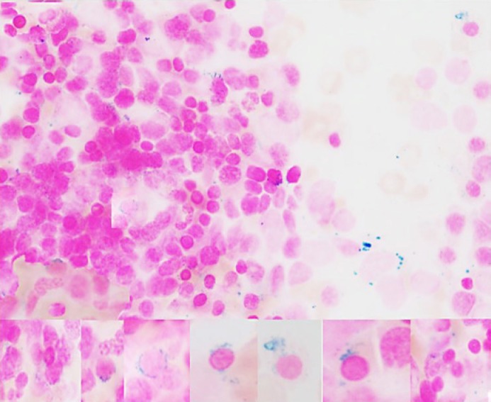
Fig. 3.
Bone marrow morphologic changes in copper deficiency. A Prominent cytoplasmic vacuolization in granulocytic precursors, eosinophils and in early erythroid precursors, left-shifted granulopoiesis with little maturation beyond the myelocyte stage, significant dysgranulopoietic features (hypo/non-segmentation and abnormal segmentation) (Wright-Giemsa. ×500). B Increased erythropoiesis with dyserythropoietic features (megaloblastoid changes, karyorrhexis, budding, binuclearity, internuclear bridging) (Wright-Giemsa. ×500).

