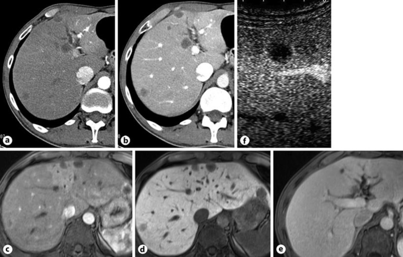
Fig. 1.
Computed tomography revealed low density tumors in both lobes of the liver (a) with ring enhancement in portal phase (b). Intrahepatic bile duct in segment 4 was dilated. Gadoxetic acid-enhanced magnetic resonance imaging identified low-intensity tumors in both lobes of the liver with ring enhancement (c), which were defective in the hepatocyte phase (d). eMRI also revealed the dilation of intrahepatic duct in segment 4. f Abdominal ultrasound with contrast enhancement showed an early ring-enhanced tumor, and its central lesion was completely defective in the postvascular phase.

