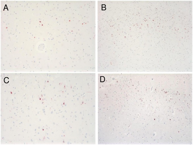
FIGURE 2.
TDP immunohistochemistry. TDP-positive granular cytoplasmic inclusions are seen in left superior temporal gyrus in Patient 1, 200× (A), in left temporal pole in Patient 2, 100× (B), in right superior temporal gyrus in Patient 3, 200× (C), and in left superior temporal gyrus in Patient 4, 100× (D). Intranuclear inclusions in all 4 patients were absent.

