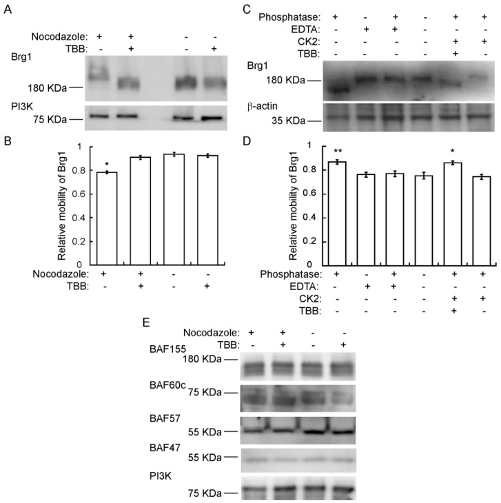
Figure 4.
Brg1 is phosphorylated by CK2 during mitosis. Primary myoblasts were treated or not with 500 nM nocodazole and 10 µM TBB, as indicated. Samples were separated in a 5% SDS-PAGE supplemented with Phos-Tag™. (A) Representative Western Blot of the CK2-mediated shift in Brg1 mobility in myoblast cultures enriched for mitotic cells. PI3K levels were monitored as a loading control. (B) Quantification of the average relative mobility of Brg1 in the gel in response to nocodazole treatment and CK2 inhibition (n = 3). (C) Representative Western Blots following an in vitro dephosphorylation/phosphorylation assay on Brg1 in primary myoblast extracts from cells treated as indicated. β-actin was monitored as a control. (D) Quantification of the average relative mobility of Brg1 in the gel in response to the treatments indicated in (C) (n = 3). (E) Representative Western Blots showing no easily discernible changes in mobility for BAF155, BAF60c, BAF57, or BAF47 in primary myoblast extracts from cells treated as indicated. PI3K was used as a loading control. Three independent biological replicates were performed for each experiment. Statistical significance was calculated by a t-test vs. the untreated control. * p < 0.05; ** p < 0.01.

