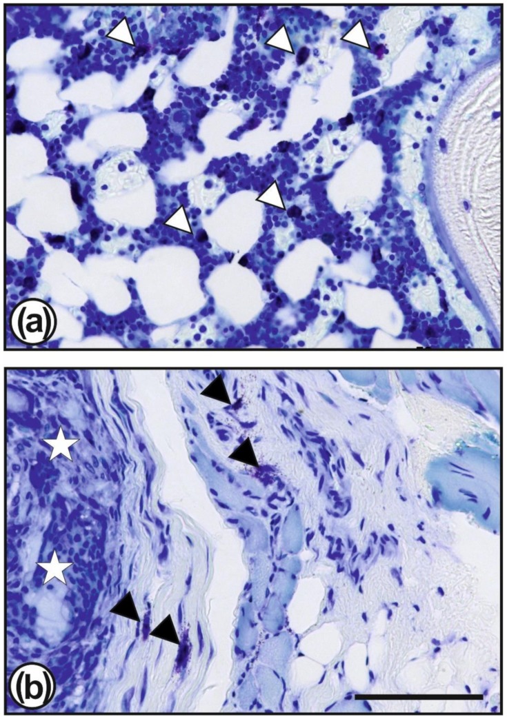
Figure 5.
Toluidine blue staining showed mast cells (some of them are marked by white or black triangles) which are present numerously in the vicinity of the surgical site, especially in the bone marrow (a) but also in the granulation tissue surrounding the TGF–β3–CS–g–PCL fibre scaffolds (b; scaffold is marked by white stars). Bar = 50 µm.

