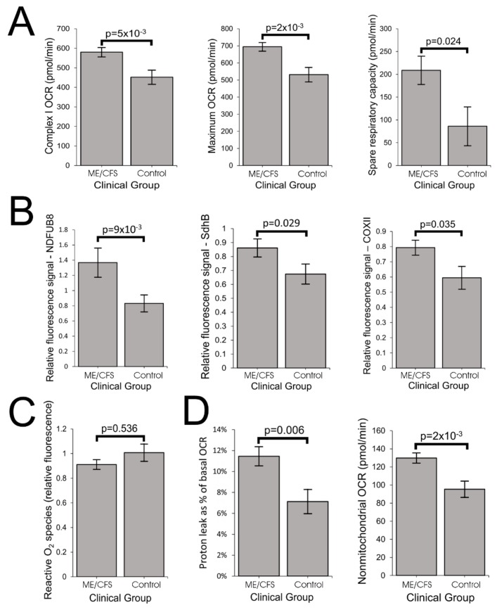
Figure 3.
ME/CFS lymphoblasts exhibit elevated respiratory capacity and expression of oxidative phosphorylation (OXPHOS) complexes. Error bars are standard errors of the mean. (A) Complex I OCR, maximum OCR and spare respiratory capacity are elevated in ME/CFS lymphoblasts (independent t-test). The OCR was measured in lymphoblasts from ME/CFS and control individuals by the Seahorse XFe24 Extracellular Flux Analyzer. Each ME/CFS (n = 50) and control (n = 22) cell line was assayed over four replicates in each of at least three independent experiments. (B) Relative expression levels of Complex I subunit NDUFB8, Complex II subunit SdhB and Complex IV subunit COXII were elevated in semiquantitative Western blots (independent t-test). Each ME/CFS (n = 48) and control (n = 17) cell line was assayed in at least three independent experiments. (C) Intracellular ROS levels (relative background-subtracted Deep Red fluorescence) are unchanged in ME/CFS lymphoblasts (independent t-test). Each ME/CFS (n = 49) and control (n = 22) cell line was assayed in duplicate within each of at least three independent experiments. (D) Proton leak as % of basal OCR and the nonmitochondrial OCR are elevated in ME/CFS lymphoblasts (independent t-test). Each ME/CFS (n = 50) and control (n = 22) cell line was assayed over four replicates per experiment in at least three independent experiments.

