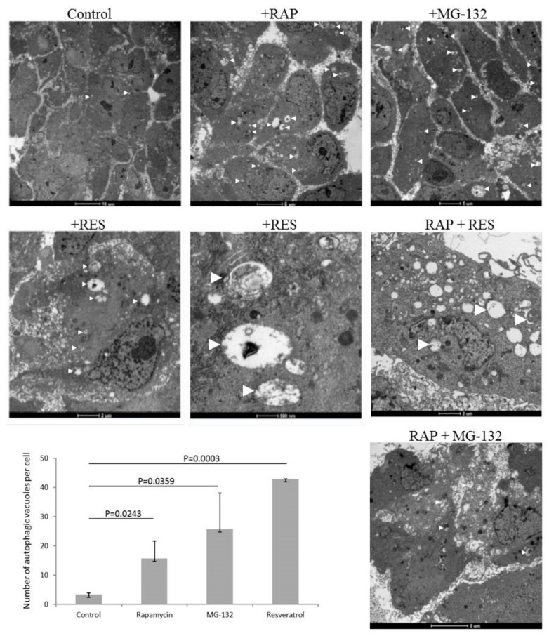
Figure 1.
ARPE-19 cell treated by autophagy inducer rapamycin (RAP, 100 nM), proteasome inhibitor MG-132 (100 nM) and resveratrol (RES, 10 µM) over 24 h. Transmission electron microscopy is shown of ARPE-19 cells under different treatment modalities. (Bars on the upper panel from left to right: 10 µm, 5 µm, and 5 µm; middle panel from left to right: 2 µm, 500 nm, and 2 µm; lower panel: 5 µm). The number of autophagic vacuoles per cell is shown for the different treatment conditions. Two-sample t-test was used for the analysis.

