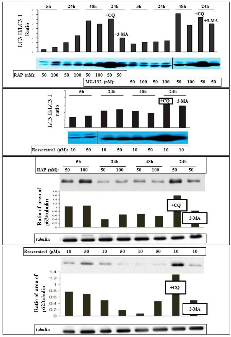
Figure 2.
Detection and quantification of autophagy in ARPE-19 cells using Western blot analysis. The cells were treated in a time- and concentration-dependent manner by the autophagy inducer rapamycin (RAP), proteasomal inhibitor MG-132, and Resveratrol. The LC3II/I ratio and expression of p62 are shown, as well as the effect of the autophagosome-lysosome fusion inhibitor chloroquine (CQ) and the upstream inhibitor of autophagy 3-methyladenine (3MA).

