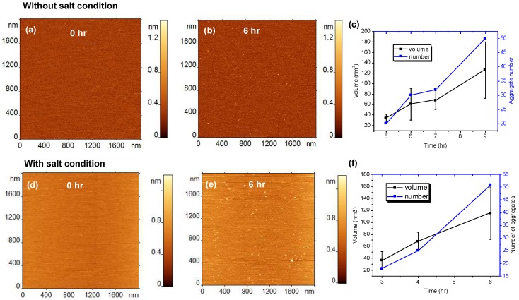
Figure 1.
Aggregation of 10 nM amyloid β 42 (Aβ42) on 1-palmitoyl-2-oleoyl-sn-glycero-3-phosphocholine (POPC) supported lipid bilayer (SLB). (a–c) Show the results obtained in the absence of 150 mM NaCl in the buffer (without salt condition) and (d–f) show the aggregation results with 150 mM NaCl (with salt condition). (a) AFM topographic image of POPC SLB just after the addition of Aβ42 onto the SLB surface. (b) The same SLB surface after incubating for 6 h, without 150 mM NaCl. The small globular features indicate the presence of aggregates. (c) The plot shows the increase in number and volume of the aggregates with time. (d–f) show similar experiments but with 150 mM NaCl added to the buffer.

