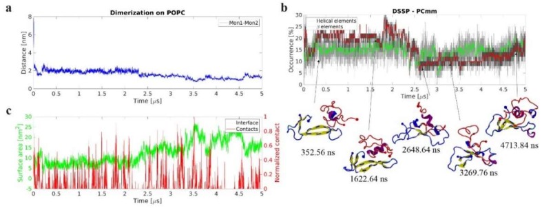
Figure 8.
Dynamics of on-surface dimerization of amyloid β42 (Aβ42) monomers in presence of 1-palmitoyl-2-oleoyl-sn-glycero-3-phosphocholine (POPC) bilayer. (a) Time-dependent center of mass (CoM) distance between the two Aβ42 monomers revealing that the dimer rapidly forms and remains stable. (b) Evolution of secondary structure β-structure (β-sheet and β-bridge, red), and helical (α-, π-, and 3/10, green), elements, as determined by DSSP. The graphs are moving averages using a 1 ns window; raw data is presented as dark and light grey graphs, respectively. Key transition points are highlighted with cartoon representation of the protein structures. Initial surface-bound monomer is depicted in blue and the initially free Aβ42 monomer in red. N- and C-terminal Cα are large and small spheres, respectively. Secondary structure elements are depicted using VMD color scheme (yellow β-strands and purple α-helices). (c) Dimer interface surface area, green, and normalized number of contacts between Aβ42 and lipid headgroups, red, showing evolution and correlation between these parameters.

