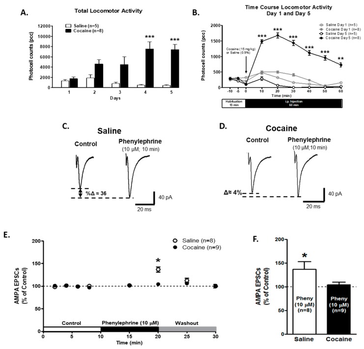
Figure 2.
α1-ARs-mediated effect on AMPA EPSCs is absent in the VTA DA neurons of cocaine sensitized animals. (A) The bar graph shows the total locomotor activity recorded from saline- (0.9%, white bars; n = 5) and cocaine- (15 mg/kg i.p., black bars; n = 8) treated animals. Cocaine-injected animals show a progressive increase in locomotor activity over 5 days. On days 4 and 5, the total activity was significantly increased when compared to day 1 in cocaine-treated animals (Two-way repeated measures ANOVA, Bonferroni post hoc test p < 0.001). (B) The time course of locomotor activity on days 1 (grey) and 5 (black) of cocaine- (15 mg/kg, i.p.) (filled) or saline- (unfilled) treated animals. Cocaine sensitization was manifested as an increase in locomotor activity during the first 10–50 min after cocaine injection in comparison with day 1. The data (pcc/60 min; Mean ± SEM) were analyzed by Two-way repeated measures ANOVA, Bonferroni post hoc test (p < 0.001). (C,D) Representative recordings from saline- and cocaine-treated animals, respectively, illustrating that phenylephrine superfusion (10 μM, 10 min) induces a significant increment in AMPA EPSCs amplitude only in VTA DA neurons from saline-treated animals. (E) A time course summary of the effects of phenylephrine bath application on AMPA EPSCs amplitude recorded from VTA DA neurons from saline-treated animals (n = 8/5) and VTA DA neurons from cocaine-treated animals (n = 9/8) at 8 min after control recording (2 min intervals), 5 and 10 min phenylephrine (10 μM), and 5 and 10 min washout. A 10 min phenylephrine application increased the AMPA EPSCs amplitude only in neurons from saline-treated animals compared to control recordings (One-way ANOVA, Newman-Keuls post-hoc, p < 0.05). A washout of phenylephrine’s response was observed. (F). A bar graph showing that, in neurons from saline-treated animals (n = 8), phenylephrine application resulted in an ~36% increase in AMPA EPSCs amplitude compared to control recordings (One-way ANOVA, Newman-Keuls post-hoc, p < 0.05). No significant differences were observed on VTA DA neurons in cocaine-treated animals (n = 9) after 10 min of phenylephrine application (104.6 ± 5.3%). Two-way repeated measures ANOVA showed in factor time, at 10 min of phenylephrine superfusion (F7,105 = 2.831, Bonferroni post hoc, p < 0.05) * p < 0.05; ** p < 0.01, *** p < 0.001. n = the number of cells recorded/number of animals.

