Figure 5.
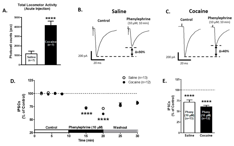
α1-ARs-mediated effect on GABAA IPSCs amplitude is not affected in VTA DA neurons after one saline or cocaine injection. (A) A bar graph showing the total locomotor activity recorded from saline- (0.9%, white bar; n = 7) and cocaine- (15 mg/kg i.p., black bar; n = 7) treated animals. The locomotion of cocaine treated animals was significantly increased compared to saline-treated controls (Unpaired t-test, p < 0.0001). (B,C) Representative recordings from saline- and cocaine-treated animals, respectively, show that phenylephrine superfusion (10 μM, 10 min) induces a significant decrease in GABAA IPSCs amplitude in VTA DA neurons from both groups. (D) The time course summary of phenylephrine effects on GABA IPSCs recorded from VTA DA neurons from saline- (n = 13) and cocaine- (n = 12) treated animals at 8 min of control (2 min intervals), 5 and 10 min phenylephrine (10 μM), and 5 and 10 min washout. A 10 min phenylephrine application decreases the GABAA IPSCs amplitude in neurons from saline- and cocaine-treated animals (One-way ANOVA, Newman-Keuls post hoc test, p < 0.0001). (E) A bar graph showing that phenylephrine application results in a ~30% decrease in GABAA IPSCs amplitude in VTA DA neurons from saline- (n = 13/7) and cocaine- (n = 12/7) treated animals (One-way ANOVA, Newman-Keuls post-hoc, p < 0.0001). **** p < 0.0001. n = the number of cells recorded/number of animals.

