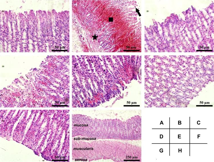
Figure 5.
Histological details of the gastric mucosa in Wistar rats. The microscopic examination was performed on the gastric mucosa obtained from different groups: (A) Control, (B) Model, (C) Sucralfate, (D) BCM, (E) L-Se, (F, H) M-Se and (G) H-Se. Briefly, Wistar rats were treated as presented in Table 1, and they were sacrificed after the ethanol challenge. The stomach was immediately obtained, part of which was quickly fixed in formaldehyde solution, routinely embedded by paraffin, sectioned (5 μm thick) and finally stained by hematoxylin and eosin (H&E) duel-dye. The Sections were examined under a light microscope. Typical gastric mucosal damage including epithelial exfoliation (arrow), mucosal hemorrhage (square) and inflammatory cell infiltration (star) was found in Model group. (A–G) magnification, × 200; (H) magnification, × 40.

