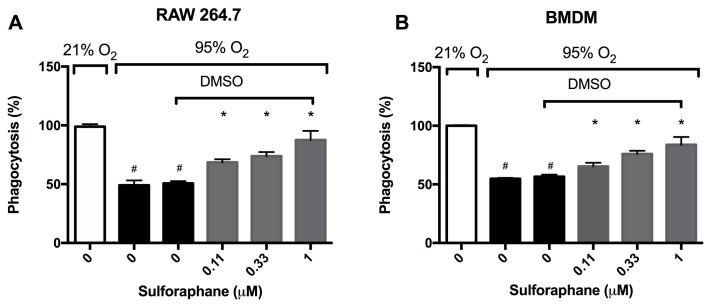
Figure 2.
Sulforaphane (SFN) attenuates the hyperoxia-induced impairment of macrophage phagocytosis. RAW 264.7 cells (A) and BMDM cells (B) were exposed to 21% O2 or 95% O2 in the presence of increasing concentrations of SFN (diluted in DMSO as the vehicle) for 24 h and were then incubated with fluorescein isothiocyanate (FITC) labeled minibeads for 1 h. Cells were subsequently stained with DAPI and phalloidin to visualize the cells. Phagocytic activity was quantified by counting the number of minibeads in at least 200 cells per well. Data are presented as the mean ± SEM of the percentage of phagocytosed minibeads. The results were based on three independent experiments. # p < 0.05 compared to 21% O2 control group. * p < 0.05 compared to 0 µM SFN vehicle control group.

