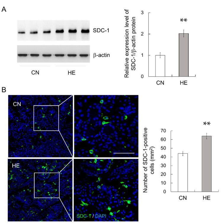
Figure 6.
Syndecan-1 (SDC-1) expression in the submandibular glands (SMGs) of control (CN) and heat-exposed (HE) rats. (A) SDC-1 protein expression in the SMGs. Heat exposure increased SDC-1 protein expression in the SMGs. Left 3 lanes show CN rats and right 3 lanes show HE rats. (B) Immunohistochemical analysis of SDC-1 (green) in the SMGs. The nuclei were stained with 4′,6-diamidino-2-phenylindole (DAPI, blue). The right panel shows magnified views of the boxed regions from the CN and HE groups. Right graph shows the density of SDC-1-immunopositive cells in the SMG sections. Values are presented as the mean ± SEM (n = 8 in each group). ** p < 0.01, significant difference between CN and HE groups. Scale bar, 25 μm.

