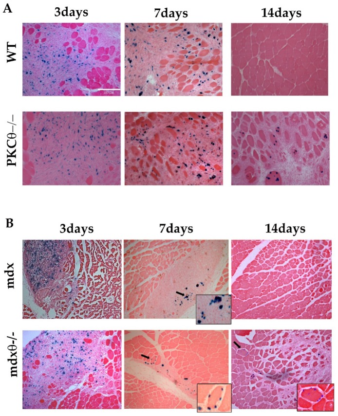
Figure 6.
(A) Representative cryosections of TA muscle derived from WT and PKCθ−/− sacrificed 3, 7, or 14 days after transplantation (n = 3–5/genotype, each time point), as indicated, and processed for X-gal activity (bar = 100µm). (B) Representative cryosections of TA muscle derived from mdx and mdxθ−/− sacrificed 3, 7 or 14 days after transplantation, (n = 3–5/genotype, each time point), as indicated, and processed for X-gal activity. Small side pictures show higher magnification of parts of the sections. Arrows indicate X-gal positive nuclei included within some fibers in PKCθ-null mice, in both non-dystrophic and dystrophic background.

