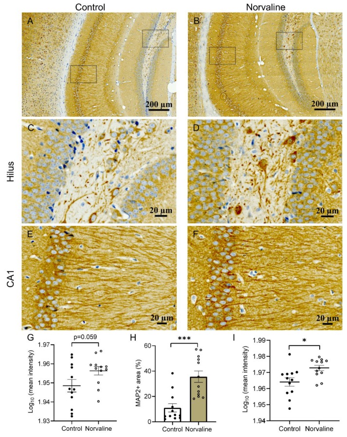
Figure 3.
Representative ×20 bright-field micrographs of the 3 × Tg mice hippocampi (A,B). ×40 magnification of hilus (C,D) and CA1 region (E,F). Norvaline treatment led to a significant increase in CA1 MAP2-immunopositive surface area (H), and stain intensity (I). (G) Mean MAP2 stain intensity in hilus area. The data are presented as means ± SEM (n = 12, four brains per group, three sections per brain). *** p < 0.001, * p < 0.05 (two-tailed Student’s t-test).

