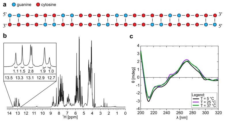
Figure 1.
1H-NMR and CD spectra of r(G2C4)4 at pH 7.0 forms dimeric structure. (a) Schematic presentation of homodimer with G-C base pairs and C-C mismatches. (b) 1H-NMR spectrum of r(G2C4)4 in 10% 2H2O at pH 7.0, 25 °C and 0.9 mM oligonucleotide concentration per strand. Numbers below the signals represent integral values. c) CD spectra of homodimer with concentration of 100 µM per strand at 5, 25 and 37 °C.

